How Muscle Contraction Works: Sliding Filament Theory
How muscle contraction works: from the nerve signal at the neuromuscular junction to actin and myosin sliding in the sarcomere, the role of calcium and ATP, and fatigue.
How muscle contraction works, in short: a nerve impulse reaches the muscle at the neuromuscular junction, the muscle fibre releases calcium ions, and these let tiny myosin heads grab the actin filaments beside them and pull, using energy from ATP. Millions of these pulls make the filaments slide past each other, every unit in the muscle shortens a little, and the whole muscle contracts. This explanation is called the sliding filament theory, put forward in 1954.
What is skeletal muscle made of?
Skeletal muscle, the kind attached to your bones, is built like a cable of smaller and smaller cables:
| Level | Size | What it is |
|---|---|---|
| Muscle | up to 50 cm | The whole muscle, such as the biceps, attached to bone by tendons |
| Fascicle | about 1 mm | A bundle of muscle fibres |
| Muscle fibre | 10–100 µm wide, up to 30 cm long | One long cell with many nuclei |
| Myofibril | 1–2 µm | Thread-like strands filling the fibre |
| Sarcomere | about 2.5 µm long | The repeating unit that actually shortens |
Each sarcomere runs from one Z-line to the next. Inside it, two protein filaments overlap:
- Actin (thin filaments), anchored to the Z-lines at each end.
- Myosin (thick filaments), in the middle, covered in tiny heads that can bind to actin.
The regular overlap of thick and thin filaments gives skeletal muscle its striped (striated) look under a microscope.
How does the nerve signal reach the muscle?
The signal to contract comes from a motor neuron. Where its axon terminal meets the muscle fibre is the neuromuscular junction, a special synapse:
- An action potential arrives at the motor neuron's axon terminal.
- Vesicles release the neurotransmitter acetylcholine (ACh) into the gap.
- ACh diffuses across and binds to receptors on the muscle fibre's membrane (the sarcolemma).
- This triggers an action potential in the muscle fibre, which spreads along it and down tubes into the fibre's interior.
- An enzyme, acetylcholinesterase, quickly breaks the ACh down, so each nerve impulse causes just one twitch.
It is the same process as a synapse between two neurons. You can watch an impulse travel down a motor neuron and neurotransmitter cross the gap in the Brain & Neurons lab, and read the full story in how a neuron works.
What is the sliding filament theory?
The sliding filament theory says that a muscle contracts because its actin and myosin filaments slide past each other, pulling the Z-lines closer together. The filaments themselves don't get shorter; they overlap more. It was proposed in 1954 by two research teams at once: Andrew Huxley and Rolf Niedergerke, and Hugh Huxley and Jean Hanson (the two Huxleys weren't related).
Step by step: the cross-bridge cycle
- Calcium is released. The muscle action potential makes the sarcoplasmic reticulum (a store inside the fibre) release calcium ions, Ca²⁺.
- Binding sites are uncovered. At rest, a protein called tropomyosin covers the binding sites on actin. Calcium binds to troponin, which moves tropomyosin out of the way.
- Cross-bridges form. Myosin heads attach to the exposed sites on actin.
- The power stroke. Each myosin head tilts, pulling the actin filament about 10 nm towards the middle of the sarcomere. ADP and phosphate are released.
- Detach and reset. A new ATP molecule binds to the myosin head, which lets go of actin. The ATP is split, and the energy re-cocks the head, ready to grab again further along.
- Relaxation. When nerve impulses stop, calcium is pumped back into the sarcoplasmic reticulum (using more ATP), tropomyosin covers the sites again and the muscle relaxes.
Each head pulls and lets go about five times a second, and the millions of heads pull at different moments, like a team hauling a rope hand over hand.
What changes in the sarcomere when it contracts?
| Part | During contraction |
|---|---|
| Sarcomere | Shorter |
| Z-lines | Closer together |
| I-band (actin only) | Shorter |
| H-zone (myosin only) | Shorter or disappears |
| A-band (length of myosin) | Stays the same |
| Actin and myosin filaments | Stay the same length |
Worked example: how much does a muscle shorten?
A sarcomere shortens from 2.5 µm to 2.0 µm when it contracts.
percentage shortening = (2.5 − 2.0) ÷ 2.5 × 100 = 20%
Every sarcomere in a fibre shortens at once, so a 30 cm muscle with this much contraction would shorten by 0.20 × 30 = 6 cm, enough to bend the elbow a long way.
Why do muscles work in pairs?
Muscles can only pull; they can't push. So skeletal muscle movement needs antagonistic pairs: when one muscle contracts, its partner relaxes and is stretched. To bend your arm the biceps contracts while the triceps relaxes; to straighten it the triceps contracts while the biceps relaxes. The brain's motor cortex and cerebellum time these pairs so movements are smooth.
Common mistakes
- Saying the filaments shorten. Actin and myosin stay the same length; they slide over each other, so the sarcomere shortens.
- Saying ATP is only needed for the power stroke. ATP is needed to detach myosin from actin, and to pump calcium back. Without ATP, muscles lock rigid (that is rigor mortis after death).
- Forgetting calcium's role. Calcium uncovers the binding sites on actin by moving troponin and tropomyosin; without it, no cross-bridges form.
- Mixing up the neuromuscular junction with a tendon. The junction is where the nerve meets the muscle; the tendon is where muscle meets bone.
Frequently asked questions
What is the role of ATP in muscle contraction?
ATP has three jobs in muscle contraction. Splitting ATP re-cocks the myosin head, storing energy for the power stroke. A fresh ATP molecule must bind before myosin can let go of actin, so the cycle can repeat. And ATP powers the pumps that return calcium to the sarcoplasmic reticulum so the muscle can relax. Without ATP, muscles stay locked, as in rigor mortis.
What happens during muscle fatigue?
Muscle fatigue is a fall in the force a muscle can produce during hard or long exercise. It happens when stores of ATP, creatine phosphate and glycogen run low, when lactic acid and phosphate build up from anaerobic respiration, and when less calcium is released from the sarcoplasmic reticulum. Rest lets the muscle restore its energy stores and clear the waste products.
How do muscles get stronger?
When you exercise a muscle against a heavy load, its fibres make more actin and myosin and more myofibrils, so each fibre gets thicker. Muscle growth comes mostly from bigger fibres, not more of them. More filaments side by side means more cross-bridges pulling at once, which means more force. Nerves also get better at activating many fibres together.
Are heart muscle and skeletal muscle the same?
Both use actin, myosin and the sliding filament mechanism, and both look striped. But heart (cardiac) muscle contracts by itself without nerve impulses, its cells are branched and joined so they beat together, and it never tires. Skeletal muscle only contracts when a motor neuron tells it to, and it does fatigue.
Try it yourself
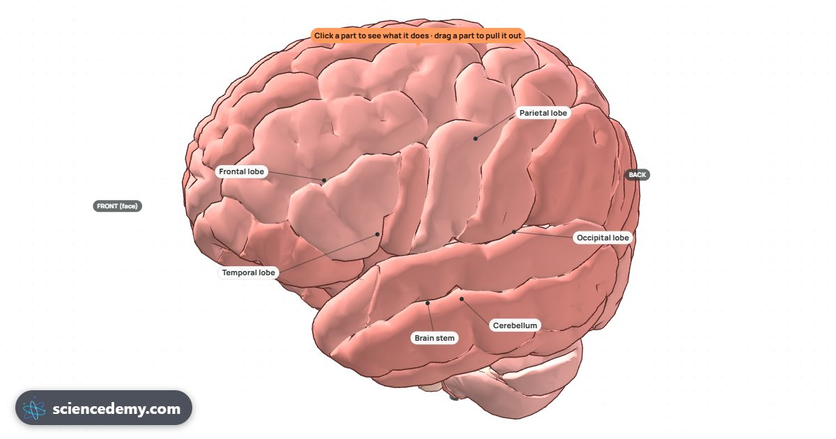
Open the Brain & Neurons lab and press ✋ Moving to watch the signal leave the motor cortex and travel down the brain stem towards the muscles. Then switch to A neuron, press Stimulate and watch neurotransmitter cross the synapse: exactly what happens at a neuromuscular junction. To see muscle power the heart, open the Heart & Circulation lab.
Voyez-le par vous-même, en 3D et gratuitement.
Essayez-le dans le Brain & Neurons