Heart Diagram (Labeled)
A labelled diagram of the heart, showing its four chambers, four valves, and the major blood vessels that carry blood in and out.
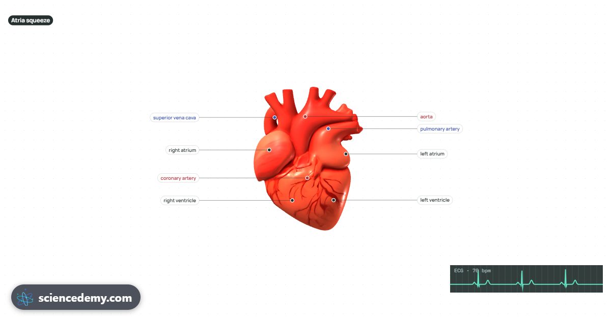
The heart's chambers and valves
Blood enters the heart through the two atria (upper chambers) and is pumped out by the two ventricles (lower chambers) below them. Between each atrium and its ventricle is a valve — tricuspid on the right, bicuspid (mitral) on the left — that snaps shut to stop blood flowing backwards as the ventricle squeezes. Two more valves, the pulmonary and aortic valves, sit where blood leaves the ventricles for the same reason.
Where the blood goes
- Vena cava — brings deoxygenated blood from the body into the right atrium.
- Pulmonary artery — carries that blood from the right ventricle to the lungs to pick up oxygen.
- Pulmonary vein — returns oxygenated blood from the lungs to the left atrium.
- الشريان الأورطي — carries oxygenated blood from the left ventricle out to the rest of the body.
This is double circulation: every drop of blood passes through the heart twice on each full circuit — once to the lungs and once to the body — which is why the heart has two separate pumps side by side, split by the muscular septum.
Open the heart in 3D below: watch it beat in time with an ECG, see the valves open and close, and follow blood through both sides of the double circulation.
الأسئلة الشائعة
What are the four chambers of the heart called?
The right atrium and left atrium (the upper chambers, which receive blood) and the right ventricle and left ventricle (the lower chambers, which pump it out).
What are the four valves of the heart?
The tricuspid and bicuspid (mitral) valves, between each atrium and ventricle, and the pulmonary and aortic valves, where blood leaves the ventricles. All four stop blood flowing backwards.
Which side of the heart pumps blood to the lungs?
The right side: the right ventricle pumps deoxygenated blood through the pulmonary artery to the lungs. The left side pumps oxygenated blood to the rest of the body through the aorta.
What is the septum?
The wall of muscle down the middle of the heart that keeps the oxygenated blood on the left side separate from the deoxygenated blood on the right side.
